What is Entropy
The GE HealthCare Entropy™ Module is indicated for adult and pediatric patients older than 2 years within a hospital for monitoring the state of the brain by data acquisition of electroencephalograph (EEG) and frontal electromyograph (FEMG) signals. The spectral entropies, Response Entropy (RE) and State Entropy (SE), are processed EEG and FEMG variables.
In adult patients, Response Entropy (RE) and State Entropy (SE) may be used as an aid in monitoring the effects of certain anesthetic agents, which may help the user to titrate anesthetic drugs according to the individual needs of adult patients. Furthermore in adults, the use of Entropy parameters may be associated with a reduction of anesthetic use and faster emergence from anesthesia.1,2,3 The Entropy measurement is to be used as an adjunct to other physiological parameters.
How is Entropy calculated
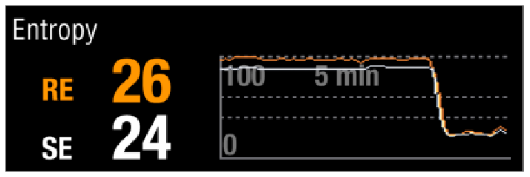
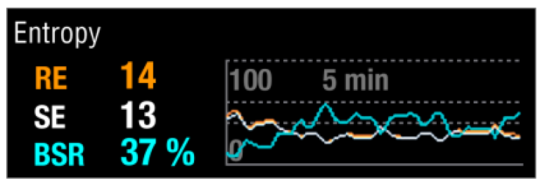
Entropy is a measure of irregularity in any signal. During general anesthesia, EEG changes from irregular to more regular patterns when anesthesia deepens. Similarly, FEMG quiets down as the deeper parts of the brain are increasingly saturated with anesthetics. The Entropy Module measures these changes by quantifying the irregularity of EEG and FEMG signals.4
In adults, the Entropy Range Guideline reflects a general association between the patient’s clinical status and Entropy values. The Guideline is based on an Entropy validation study5 involving administration of specific anesthetic agents. Titration of anesthetics to Entropy Guideline should be done in context of patient status and treatment plan. Individual patients may show different values.
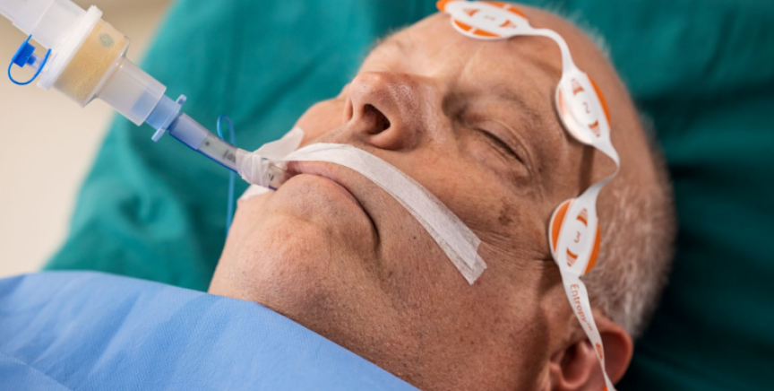
Getting started
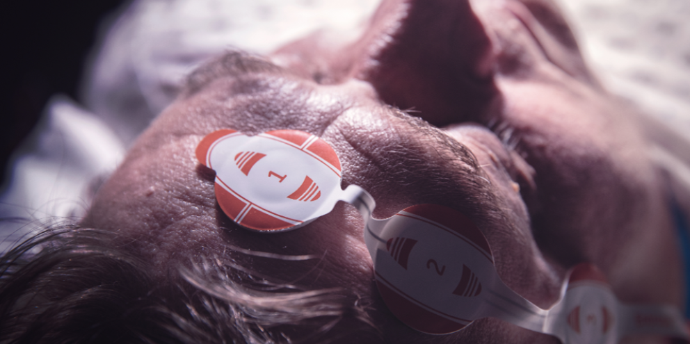
Monitoring the electrical activity of the brain and facial muscles with the Entropy Module is easy, just attach the Entropy sensor on the patient’s forehead according to the instructions provided on the sensor pouch. The module automatically checks that the electrode impedances are within an acceptable range and starts the measurement. The measurement will continue until the sensor is removed.
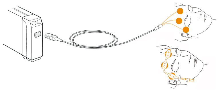
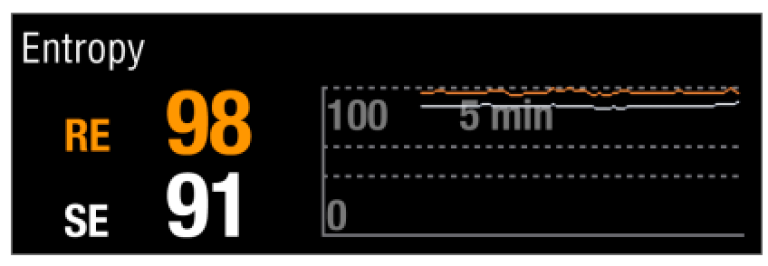
In adults, Entropy values have been shown to correlate to the patient’s anesthetic state. High values of Entropy indicate high irregularity of the signal, signifying that the patient is awake. A more regular signal produces low Entropy values which can be associated with low probability of consciousness.
Entropy Range Guidelines
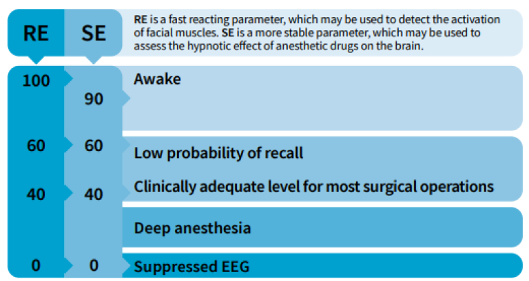
* Frequent eye movements, coughing and patient movement cause artifacts and may interfere with the measurement. Entropy readings may be inconsistent when monitoring patients with neurological disorders, traumas or their sequelae. Epileptic seizures and psychoactive medication may cause inconsistent Entropy readings.
The Entropy parameters
There are two Entropy parameters: the fast-reacting Response Entropy (RE) and the more steady and robust State Entropy (SE). State Entropy consists of the entropy of the EEG signal calculated up to 32 Hz. Response Entropy includes additional high frequencies up to 47 Hz. Consequently the fast frontalis EMG (FEMG) signals enable a fast response time for RE.

Response Entropy (display range 0-100)
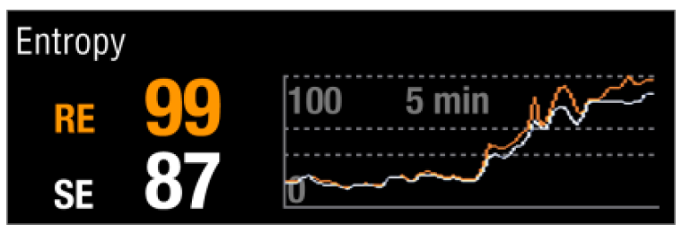
Response Entropy is sensitive to the activation of facial muscles, (i.e., FEMG). Its response time is very fast; less than 2 seconds. FEMG is especially active during the awake state but may also activate during surgery. Facial muscles may also give an early indication of emergence, and this can be seen as a quick rise in RE.
State Entropy (display range 0 - 91)
The State Entropy value is always less than or equal to Response Entropy. During general anesthesia the hypnotic effect of certain anesthetic drugs on the brain may be estimated by State Entropy value. SE is less affected by sudden reactions to the facial muscles because it is mostly based on the EEG signal.
Neuromuscular blocking agents (NMBA), administered in surgically appropriate doses are not known to affect the EEG, but are known to have an effect on the EMG.

Why use Entropy monitoring
Use Entropy to titrate anesthetic drugs according to the individual needs of the patient
Entropy measures the activity of the brain, which is the target organ for anesthetic medication, and has been shown to reflect the different phases of anesthesia. Furthermore, the use of Entropy has been found to reduce the use of certain hypnotic drugs. Therefore, Entropy may help you tailor the administration of anesthetic drugs for each patient individually.
Use Entropy to control recovery and to improve your perioperative process
With Entropy monitoring, it is possible to ensure faster emergence and recovery in the operating room. It is a tool for optimizing the perioperative process and ensuring efficient patient flow.
Integrated information
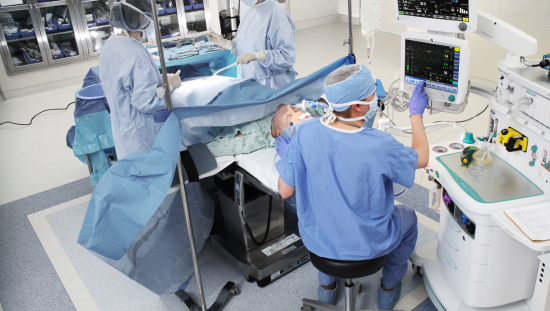
When Entropy monitoring is integrated into a monitoring system, the measured values are displayed, trended, and automatically documented together with all of the other monitored parameters.
Adequacy of Anesthesia
Unconsciousness, amnesia, antinociception and at least some degree of immobility combined with autonomic stability are various targets, when tailoring anesthesia for each patient. Therefore, it takes more than one measurement to reliably assess the adequacy of anesthesia. With Entropy used together with other monitored parameters, such as the hemodynamic measurements and NMT, you can get a complete picture of the patient status combined on one screen. All these values are stored in the monitor memory for trending and information management purposes.
At GE HealthCare, our vision is to provide you with the complete range of clinical parameters to help you give personalized anesthesia for each patient.
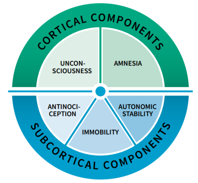
Clinical use of Entropy


Additional resources
For white papers, guides and other instructive materials about our clinical measurements, technologies and applications, please visit http://clinicalview.gehealthcare.com/
References
- Vakkuri et al. Spectral entropy monitoring is associated with reduced propofol use and faster emergence in propofol - nitrous oxide - alfentanil anesthesia. Anesthesiology 103, 274-9 (2005).
- 2. Aimé, I., et al. Does monitoring bispectral index or spectral entropy reduce sevoflurane use? Anesthesia and Analgesia, 103(6), 1469–1477 (2006).
- 3. El Hor, T., et al. Impact of entropy monitoring on volatile anesthetic uptake. Anesthesiology, 118(4), 868–873 (2013).
- 4. Viertiö-Oja et al. Description of the Entropy algorithm as applied in the Datex-Ohmeda S/5 Entropy Module. Acta Anaesthesiologica Scandinavica 48, Issue 2: 154-161 (2004).
- 5. Vakkuri et al. Time-frequency balanced spectral entropy as a measure of anesthetic drug effect in central nervous system during sevoflurane, propofol, and thiopental anesthesia. Acta Anaesthesiologica Scandinavica 48, Issue 2: 145-153 (2004).
- 6. Klockars et al. Spectral entropy as a measure of hypnosis in children. Anesthesiology 104, 708-17 (2006).

