A patient’s VO2 is determined by oxygen delivery (DO2)—the product of cardiac output and the oxygen content in arterial blood—and oxygen extraction. Lower VO2 is associated with increased mortality risk in patients with sepsis3, and impairment in oxygen extraction due to alteration of mitochondrial function is a major factor in both sepsis and the post-arrest state. However, the variability from patient to patient is poorly understood.4-7
A GE HealthCare CARESCAPE™ monitor with a gas module with metabolics functionality has made VO2, carbon dioxide production (VCO2), and the respiratory quotient (RQ) more feasible in the intensive care setting.
Design and setting
Uber and colleagues conducted a single-center observational study of trends in gas metabolism in patients after cardiac arrest. The researchers hypothesized that there would be abnormalities in metabolism after cardiac arrest, and that lower VO2, VCO2, and RQ would be associated with lactate and clinical outcomes.
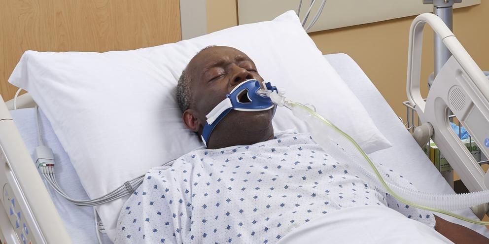
The study was conducted at an urban tertiary care center in Boston from January 2016 through July 2017. Patients were part of an ongoing prospective observational study of metabolism in critically ill adults receiving invasive mechanical ventilation. To minimize variation in temperature and sedation (both of which can alter VO2), the study population was limited to patients who received therapeutic temperature management (TTM) after in-hospital or out-of-hospital cardiac arrest, with a body temperature ≤36°C at the time of data collection.
Also excluded from the study were patients who (1) exhibited factors known to alter VO2 measurement, such as agitation or known air leak; (2) had a positive end-expiratory pressure (PEEP) >12 cm H2O, as connecting the monitor requires a brief disconnection from the ventilator circuit; (3) required fraction of inspired oxygen (FiO2) >60%, given the requirements of the monitoring technology (described below), or (4) were enrolled concurrently in an ongoing randomized trial of Ubiquinol after cardiac arrest, due to the theoretical effect of Ubiquinol on VO2.
VO2 technology
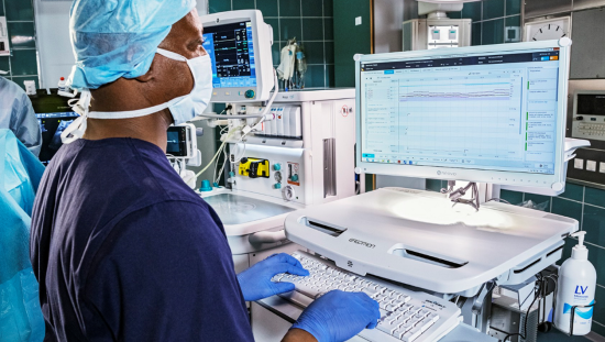
Continuous VO2 and VCO2 measurements were obtained using the CARESCAPE™ Monitor B650 with CARESCAPE Respiratory Module E-sCOVX (GE HealthCare). Values were recorded and saved, once per minute, using the accompanying GE HealthCare S/5 Collect software. RQ values were calculated by dividing VCO2 by VO2. The researchers included all values when measurements appeared to represent steady state (not changing by more than ~10% over several minutes).
The CARESCAPE monitor connects in-line with the patient’s ventilator tubing and has a built-in module for measuring spirometry and gas exchange. Via gas-sampling ports and a flow sensor, the module measures the flow of exhaled gas and the difference in oxygen and carbon dioxide content between inhalation and exhalation, using a pneumotachograph and a rapid paramagnetic analyzer. The S/5 Collect software records all values averaged over the chosen interval (e.g., every minute). This monitor has been cleared to measure VO2 and VCO2 in critically ill, mechanically ventilated patients and has been validated against indirect calorimetry using the metabolic cart. For information on limitations of VO2 monitoring, please refer to the CARESCAPE monitors User Manuals.
Results
Of the 78 patients enrolled initially, 23 were post-cardiac arrest and had data collected during TTM. Six of the latter were excluded because of enrollment in the Ubiquinol study, leaving 17 patients for analysis. Among this cohort of 17, VO2 in the first 12 hours after return of spontaneous circulation (ROSC) was associated with survival (median for survivors: 3.35 mL/kg/min [2.98,3.88]; median for non-survivors: 2.61 mL/kg/min [2.21,2.94]; p = .039). However, this did not persist through 24 hours. The ratio of VO2 to lactate correlated with survival (median for survivors; 1.4 [IQR: 1.1,1.7]; median for non-survivors, 0.8 [IQR: 0.6,1.2]; p < .001). Median RQ for the full cohort was 0.66 (IQR: 0.63,0.70), and 71% of all RQ measurements were below 0.7. Patients whose initial RQ was below 0.7 had a survival rate of 17%, whereas those whose initial RQ exceeded 0.7 had a survival rate of 64% (p = .131). No correlation was observed between VCO2 data and the probability of survival.
Conclusions
Although Uber and colleagues found a significant link between VO2 and mortality in the first 12 hours following ROSC, this association did not persist through 24 hours. Lower ratios of VO2 to lactate were associated with greater risk of mortality. There was no significant difference in RQ over 12 or 24 hours. Further research is needed to explore whether these parameters could have true prognostic value or become potential targets for treatment.
References
- Ayoub IM, Radhakrishnan J, Gazmuri RJ. Targeting mitochondria for resuscitation from cardiac arrest. Crit Care Med. 2008;36:S440–6. Accessed May 3, 2019.
- Uber et al. Preliminary observations in systemic oxygen consumption during targeted temperature management after cardiac arrest. Resuscitation. 2018;127:89-94. Accessed May 3, 2019.
- Dunn, J. O., Mythen, M. G., & Grocott, M. P. (2016). Physiology of oxygen transport. Bja Education, 16(10), 341-348.
- Fink MP. Bench-to-bedside review: cytopathic hypoxia. Crit Care. 2002;6:491–499. Accessed May 3, 2019.
- Vandermeer TJ, Wang H, Fink MP. Endotoxemia causes ileal mucosal acidosis in the absence of mucosal hypoxia in a normodynamic porcine model of septic shock. Crit Care Med. 1995;23:1217–1226. Accessed May 3, 2019.
- Rosser DM, Stidwill RP, Jacobson D, Singer M. Oxygen tension in the bladder epithelium rises in both high and low cardiac output endotoxemic sepsis. J Appl Physiol. 1995;79:1878–1882. Accessed May 3, 2019.
- Astiz M, Rackow EC, Weil MH, Schumer W. Early impairment of oxidative metabolism and energy production in severe sepsis. Circ Shock. 1988;26:311–320. Accessed May 3, 2019.

