Neuromuscular monitoring and residual paralysis
Before and during surgery, neuromuscular-blocking agents (NMBAs) are used to facilitate intubation and ensure optimal conditions for surgery. However, patients who receive NMBAs may be at a risk for adverse postoperative effects of these agents. Residual paralysis is a major concern and can lead to respiratory complications including an increased risk of aspiration and pneumonia, hypoxemia, airway obstruction, the need for tracheal intubation, and increased length of stay in the postanesthesia care unit (PACU) or potentially admission to the ICU in case of complications.1
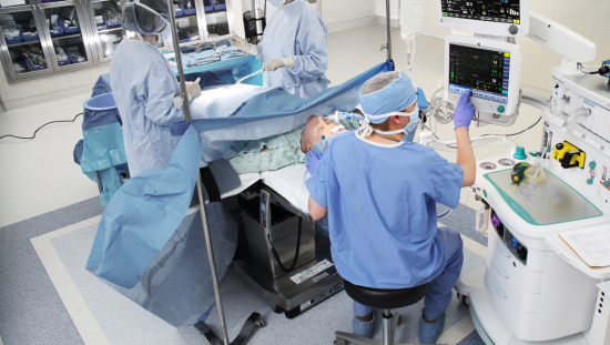
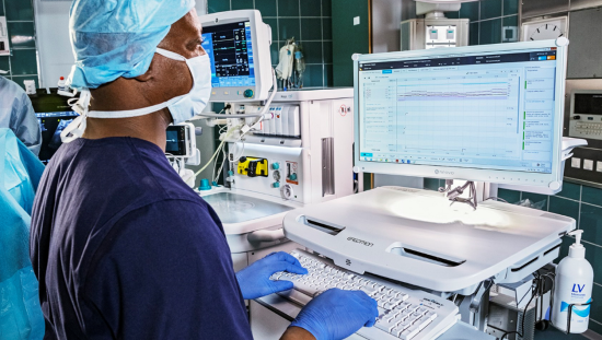
During anesthesia administration, the anesthetist must continually assess a patient’s neuromuscular function. Typical clinical observations for such include the evaluation of grip strength and head lift; however, these techniques are unreliable and difficult to use in anesthesia-emergent patients. Separately, qualitative peripheral nerve stimulation assesses the response of the stimulated muscle. Unfortunately, this technique does not eliminate the risk of residual paralysis. As such, specialized quantitative monitors are available to supplement the above options. Those neuromuscular transmission (NMT) monitors provide automatic, numerical measurements indicating the muscle response to a stimulus and the associated level of neuromuscular block.
The myths and misperceptions of NMT monitoring
Research indicates, however, that the use of NMT monitoring devices is highly variable among anesthesia practitioners as well as between countries and institutions.2,3 In certain countries, such as Finland, NMT is considered the standard of care and is required whenever an NMBA is used. Likewise, the American Society of Anesthesiologists (ASA), the European Society of Anaesthesiology and Intensive Care (ESAIC), as well as the Anesthesia Patient Safety Foundation (APSF) independently came to similarly strong recommendations to use quantitative neuromuscular monitoring.
According to Laitinen and Sarkela, there are various reasons for why practitioners or institutions may not require the use of NMT, and some perceived issues relate to the monitoring devices themselves.
“Even if there is [a] willingness to use the devices, they are simply not available at every institution,” remarks Laitinen.
Myth 1: Visual and tactile assessment suffices
One widespread myth preventing ubiquitous NMT use is the belief that it is possible to observe a patient’s NMB status by way of visual and tactile measures alone. In some regions or particular institutions, NMT is not considered mandatory, and clinicians may instead rely only on clinical observation. “This is one of the biggest misconceptions,” said Laitinen, “and it is something we are working to change. The [clinical] literature makes it clear that this is not true,” Laitinen stressed. “Still, many clinicians think it is possible to determine the level of NMB without quantitative monitoring.”
Myth 2: Residual paralysis isn’t a significant concern
Similarly, the connection between NMB and postoperative complications may not be sufficiently recognized. Specifically, there’s a misconception that residual paralysis isn’t much of a concern. Problems may manifest only after
the release from PACU to general ward and the anesthesia personnel have no knowledge of how their patients are doing there. “Especially if an institution’s operating room is independent from the rest of the hospital, this linkage [between the lack of NMT and rates of residual paralysis] may not be clear,” Laitinen explained.
Myth 3: NMT monitoring is complex and unreliable
Some clinicians view the setup and sensor placement required for measurement as complex and time-consuming. Some may also view certain devices as unreliable or prone to errors. Furthermore, components can be misplaced, as most NMT devices are handheld pieces of equipment that do not integrate into the monitoring system, and availability is a problem as well. Though previous technologies may have seemed complicated and difficult to learn, new advances and NMT monitoring devices have made monitoring easier, more reliable, and less complex.
Myth 4: The lack of official support for NMT
Another obstacle is the perceived lack of official support for NMT use. Clinicians may feel as though there aren’t official recommendations offered for NMT monitoring. However, professional societies such as the European Society of Anaesthesiology (ESAIC), the American Society of Anesthesiologists (ASA) and the Anesthesia Patient Safety Foundation (APSF) have delivered clear and strong guidelines related to quantitative monitoring. 4,5,6
NMB reversal
At the conclusion of surgery, agents are frequently used to reverse NMB. The main classes of reversal agents are anticholinesterases (e.g., neostigmine) and cyclodextrins (e.g., sugammadex). The choice of reversal agent depends on many factors, including the NMBA in use, the profoundness of the blockade, the time available for reversal, and cost considerations.
The most commonly employed nondepolarizing NMB agents are rocuronium bromide and vecuronium bromide. Both neostigmine and sugammadex may be used to reverse blockage achieved with either of these NMB agents.
Neostigmine
Neostigmine can reverse the effects of many commonly used NMBAs, including atracurium and cisatracurium. However, the time needed to achieve adequate reversal with this drug may vary significantly between patients. Moreover, neostigmine is efficient only when the NMB has spontaneously returned to one or two twitches on train-of-four (TOF) monitoring. Neostigmine will not reverse deep blockade.
Sugammadex
Sugammadex is effective only for reversing the effects of rocuronium or vecuronium, although it can be successful in reducing very deep blockade. “Many consider sugammadex a ‘miracle drug,’” said Laitinen. “It is very efficient, has few side effects, and does the job with all patient groups and in all levels of paralysis.” Because of these advantages, some may feel sugammadex does not require quantitative monitoring. However, too-early administration or a too-small dosage of sugammadex may lead to residual paralysis. In addition, depending on the geographic region, sugammadex may cost up to 20 times more than neostigmine, and many institutions have strict protocols for the use of this agent because of this prohibitive cost.
Regardless of the agent chosen to reverse NMB, none of these drugs preclude the need for quantitative NMT monitoring. “In fact, when both neostigmine and sugammadex are options, NMT may help [in deciding] between the two,” Laitinen suggested. NMT can also influence dosage size and timing of drug administration. For example, sugammadex is a very dose-dependent drug; the amount required to reverse a deep blockade may be four to eight times larger than that needed to mitigate a very light blockade. Underdosing increases the risk of residual paralysis. “It is very important to know how deep the blockade is in order to know how much sugammadex to administer,” Laitinen explained.
Measuring neuromuscular blockade
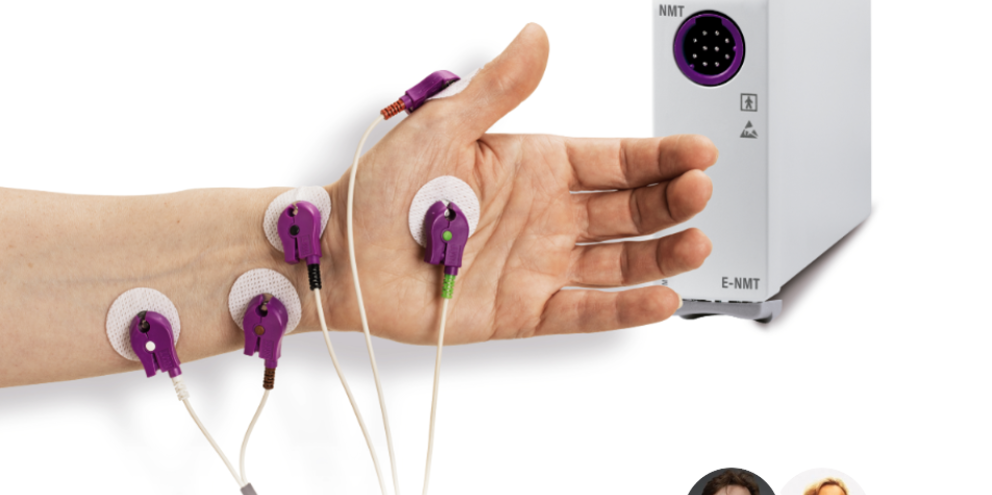
Clinicians routinely measure the depth of NMB using electrodes and a small current to stimulate peripheral nerves, most commonly the ulnar nerve in the hand, or the medial plantar nerve in the foot if the former is unavailable. Muscle response is automatically measured with a device. This so-called TOF monitoring assesses neuromuscular transmission based on the number of muscle responses detected. The TOF ratio is the ratio of the fourth muscle response to the first one and indicates NMB fade with non-depolarizing NMBAs. Extubation after surgery is recommended only if the TOF ratio exceeds 0.9.1
Currently available NMT monitoring technology includes electromyography (EMG), which measures muscle fiber potentials; kinemyography (KMG), which measures thumb bending caused by the stimulus; and acceleromyography (AMG), which measures thumb acceleration.
In EMG NMT monitoring, clinicians measure the depolarization of the muscle membrane close to the site of the NMB action and directly measures electrical activity. Considered the gold standard (besides MMG, which is mainly used in the research environment), EMG measurement does not require any physical movement of the hand. “It works well even if the hand is not available at all or completely immobilized for the operation,” noted Sarkela, who emphasized that devices for measuring KMG and AMG in comparison have limitations, adding that “[their] quality of the signal is not as good as EMG.” For example, a TOF ratio of 0.9 measured with an AMG-based NMT monitoring device may not indicate the achievement of adequate recovery for safe extubation. “There is [a] growing awareness of the superiority of the EMG signal1,” Laitinen added.
The GE HealthCare solution for NMT monitoring
GE HealthCare offers a fully integrated NMT measurement module with the ability to measure both KMG- and EMG-based signals. The following three different NMT sensors provide maximal flexibility: the MechanoSensor (KMG), pediatric MechanoSensor (KMG), and ElectroSensor (EMG).
Concluding remarks
Residual paralysis from anesthesia following surgery is a common problem despite the use of reversal agents. According to the literature, 20% to 40% of patients arrive in the PACU with signs of residual paralysis.7
Laitinen and Sarkela concur that this issue relates to the lack of quantitative NMT monitoring when applying reversal agents. Prior surveys also concur, indicating that objective paralysis monitoring is performed in only 17% of patients.7
During the discussion, Laitinen commented that “the most proven and important benefits of NMT monitoring are to optimize NMBA dosage and, crucially, from a patient’s point of view, to be able to predict recovery and avoid residual paralysis.”
Resources:
- Duţu M, Ivaşcu R, Tudorache O, et al. Neuromuscular monitoring: an update. Rom J Anaesth Intensive Care. 2018;25(1):55–60. DOI: 10.21454/rjaic.7518.251.nrm. Accessed July 4, 2019.
- Hammermeister KE, Bronsert M, Richman JS, Henderson WG. Residual neuromuscular blockade (NMB), reversal, and perioperative outcomes. Anesthesia Patient Safety Foundation (APSF) Newsletter. 2016;30(3):45–76. Accessed July 4, 2019
- Checketts MR, Alladi R, Ferguson K, et al. Recommendations for standards of monitoring during anaesthesia and recovery 2015: Association of Anaesthetists of Great Britain and Ireland. Anaesthesia. 2016;71(1):85–93. DOI: 10.1111/anae.13316. Accessed July 4, 2019
- Stephan R. Thilen, Wade A. Weigel, Michael M. Todd, Richard P. Dutton, Cynthia A. Lien, Stuart A. Grant, Joseph W. Szokol, Lars I. Eriksson, Myron Yaster, Mark D. Grant, Madhulika Agarkar, Anne M. Marbella, Jaime F. Blanck, Karen B. Domino; 2023 American Society of Anesthesiologists Practice Guidelines for Monitoring and Antagonism of Neuromuscular Blockade: A Report by the American Society of Anesthesiologists Task Force on Neuromuscular Blockade. Anesthesiology 2023; 138:13–41 doi: https://doi.org/10.1097/ALN.0000000000004379
- Fuchs-Buder, Thomas; Romero, Carolina S.; Lewald, Heidrun; Lamperti, Massimo; Afshari, Arash; Hristovska, Ana-Marjia; Schmartz, Denis; Hinkelbein, Jochen; Longrois, Dan; Popp, Maria; de Boer, Hans D.; Sorbello, Massimiliano; Jankovic, Radmilo; Kranke, Peter. Peri-operative management of neuromuscular blockade: A guideline from the European Society of Anaesthesiology and Intensive Care. European Journal of Anaesthesiology 40(2):p 82-94, February 2023. | DOI: 10.1097/EJA.0000000000001769
- Chung, C., et al. New practice guidelines for neuromuscular blockade. Anesthesia Patient Safety Foundation. 2023 https://www.apsf.org/article/new-practice-guidelines-for-neuromuscular-….
- Brull SJ, Kopman AF. Current status of neuromuscular reversal and monitoring: challenges and opportunities. Anesthesiology. 2017;126(1):173–190. DOI: 10.1097/ALN.0000000000001409. Accessed July 4, 2019.

Mika Sarkela
Principal Engineer of neurological monitoring

Matti Laitinen
Global Product Manager, Neurology and Advanced Hemodynamics

