Causes of QT prolongation
Prolongation of the QT interval is attributed to the abnormal structure or function of ion channels and related proteins responsible for cardiac cellular repolarization.2 In the case of congenital long QT syndrome (LQTS), gene mutations are responsible for these abnormalities. Conversely, with acquired LQTS, these abnormalities are the result of acute medical conditions (e.g., hypokalemia, hypomagnesemia, hypothyroidism) or the action of certain medications.
Researchers have found that some instances of medication-induced acquired LQTS and TdP may be related to previously undetected genetic factors.2 CredibleMeds®, an independent nonprofit organization and reliable resource for clinicians across multiple areas of medicine, maintains an updated list of medications that are known to prolong the QT interval on their website. Antidepressants, antibiotics, antipsychotics and antiarrhythmics are just a few of the common classes of drugs known to cause Long QT intervals.
Methods of corrected QT interval monitoring
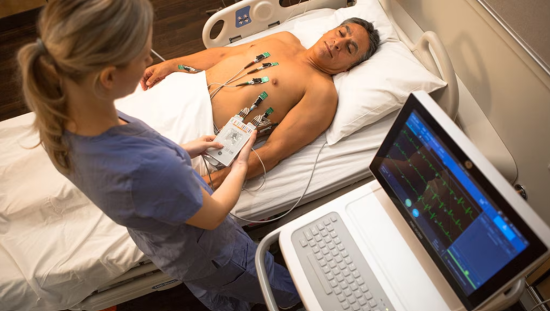
On the electrocardiogram (ECG), the QT interval begins with the onset of the QRS complex, which reflects ventricular depolarization, and ends at the T-wave, which reflects the last part of ventricular repolarization. For in-hospital patient monitoring, methods used to measure the QT and correction of the QT interval for heart rate (QTc) are generally determined by way of either (1) manually using handheld calipers, (2) using a semiautomated approach with electronic calipers built into an ECG monitoring system, or (3) fully automated continuous QTc monitoring.2
Indications for continuous corrected QT interval surveillance
In the Neonatal Intensive Care Unit (NICU)
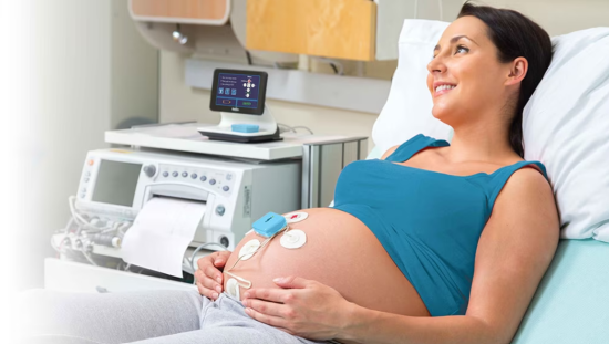
Given the fact that approximately one in 10 cases of sudden infant death syndrome can be traced back to genetic causes of prolonged QT intervals, implementing continuous monitoring solutions in the NICU could stave off the risk of medication-based QT prolongation in individuals where risks of congenital LQTS may already exist.2
After drug administration
Additionally, when a prolonged QTc interval coincides with drug administration, the existing guidelines recommend more frequent QT surveillance be performed, which represents a prime opportunity for continuous monitoring. Such indications may include patients at risk for prolonged QTc intervals before initiating nonantiarrhythmic medications with a known risk for promoting drug-induced LQTS such as amiodarone, flecainide, or dronedarone.2
Hereditary long QT
Other clinical scenarios may benefit from including continuous surveillance in certain circumstances. For example, in the case of hereditary LQTS, the AHA’s guidelines recommend QTc interval monitoring be performed until certain thresholds are met, including when (1) the QTc interval returns to a normal reading, (2) the ventricular arrhythmia is stabilized, and (3) any aggravating metabolic or health conditions are corrected.2 For these and other similar scenarios, automatic monitoring may help to indicate when the necessary thresholds are met. Automated monitoring also offers repeatable, reliable monitoring in many other diverse circumstances where manual monitoring—though valuable diagnostically—may fall short.3
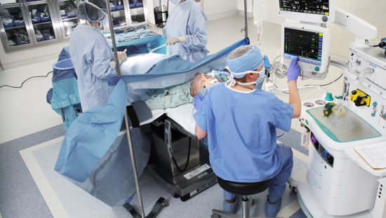
In addition, automatic QT surveillance has shown promise in perioperative care thanks to T-wave detection provided by vector magnitude signals from eight-lead inputs. Such methodology offers the benefits of being readily available, eliminating interobserver variables, and representing tested validations across nonsurgical scenarios.3
How GE HealthCare enables continuous monitoring
For each of these indications, GE HealthCare’s CARESCAPE™ monitoring system offers comprehensive, adaptable options that automate once-manual monitoring efforts for more rapid and accurate QT surveillance.4
The CARESCAPE Monitor B850, for example, consolidates and integrates multisource inputs for a cohesive data flow—all within a user interface that prioritizes ease of use. Additionally, the NICU software package enables the availability of rapid reconfigurations, alarm management control, and event annotations that feed into a beat-by-beat analysis of cardiac events.4
Such components take place within an end-to-end point-of-care context, enabling full continuity of data flow with uninterrupted remote access to manage monitoring wherever and whenever it is indicated, across multiple stages of care.4
These built-in flexibilities are what make abiding by the AHA’s guidelines more feasible at any time and place, without the need for labor-intensive and potentially inaccurate manual monitoring. Given those considerations, it’s apparent that, when appropriate per the AHA’s clinical recommendations, QT monitoring enabled by continuous surveillance is a beneficial choice for both patient outcomes and the operational management of data flow.
In addition to CARESCAPE, GE Marquette™ 12SL ECG analysis program tracks QT measurements by participating in studies to determine drugs’ QT prolongation and TdP risk, according to Ian Rowlandson, GE HealthCare Chief Engineer and Scientist for Diagnostic Electrocardiography. With the ability to assess these risks with a high degree of accuracy, clinicians can more efficiently act to reverse the effects of Long QT.
References
- Rautaharju, PM, SurawiczB, Gettes LS. AHA/ACCF/HRS Recommendations for the standardization and interpretation of the electrocardiogram: Part IV: The ST segment, T and U waves, and the QT Interval: a scientific statement from the American Heart Association Electrocardiography and Arrhythmias Committee, Council on Clinical Cardiology; the American College of Cardiology Foundation; and the Heart Rhythm Society. Endorsed by the International Society for Computerized Electrocardiology. J Am Coll Cardiol. 2009;53(11): 982–991. DOI: 10.1016/j.jacc.2008.12.014.Accessed May 10, 2019.
- Sandau, KE, FunkM, AuerbachA, et al. Update to practice standards for electrocardiographic monitoring in hospital settings: a scientific statement from the American Heart Association. Circulation. 2017;136(19):e273–e344. DOI: 10.1161/CIR.0000000000000527. Accessed May 10, 2019.
- Friederich P. Monitoring of the QTinterval in perioperative and critical care. Available at: https://www.gehealthcare.com/en/white-paper/automated-qt-interval-monit…. Accessed May 21, 2019.
- GE HealthCare. CARESCAPE Monitor B650. Available at: https://www.gehealthcare.com//-/jssmedia/6ab3979a770840cca6d2edbad89ff7…. Accessed May 21, 2019.

