Consequences of PORC include1:
- Hypoxia
- Respiratory failure
- Anoxic organ damage
- Psychomotor agitation
- Post-traumatic stress disorder
PORC is identified by a train-of-four ratio (TOFR) less than 0.9, muscle fatigue, reduction of airway protective reflexes, or a reduced rib cage expansion.[1] Once a patient has a TOFR greater than 0.9, they are felt to be safe for tracheal extubation.
Neuromuscular Blockage Monitoring
When the effects of NMBAs, or the neuromuscular blockage, are monitored, clinicians are better able to manage NMBAs and antagonists as well as establish the right time to reverse and extubate and hopefully prevent PORC events.
Monitoring the neuromuscular blockage leads to a decrease in the incidence of PORC and postoperative pulmonary complications including persistent atelectasis and aspiration pneumonitis – both of which are known to increase 30-day mortality rate in postoperative patients.[2] Postoperative pulmonary complications also increase length of hospital stay and increase overall healthcare costs.[3]
Despite the known risks of PORC and the evidence showing reduction of PORC with proper monitoring, studies show only 17% of physicians use monitoring often. 83% reported using no monitoring at all or only using subjective criteria.2
Studies have shown subjective measures using visual or tactile means don’t allow for consistent monitoring and PORC detection, while objective (quantitative) measures have been shown to improve patient safety and outcome.2 Assessing a patient’s ability to lift their head or judging the grip of a handshake have been proven to be unreliable and impossible to gauge consistently when attempting to evaluate adequate recovery.
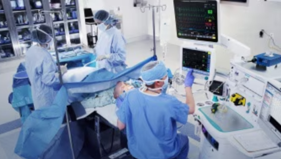
Proper neuromuscular monitoring is key to reducing complications and thankfully there are several types of objective neuromuscular monitoring technology available for clinicians to use in the operating room.
Types of NMT Technology
NMDAs do not interfere with muscle contractility or muscle membrane excitability, creating an opportunity to objectively measure neuromuscular response. There are four major neuromuscular monitoring techniques (NMT) that measure muscle acceleration, muscle contraction, the force of muscle contraction, or muscle depolarization.
Mechanomyography (MMG)
Mechanomyography (MMG) measures isometric muscle contraction caused by ulnar nerve stimulation. Although MMG measurements are highly precise and repeatable, there are several limitations to MMG technology.
MMG requires large equipment in an already pressed for space operating suite, uninterrupted access to the monitored hand, and requires the application and maintenance of pretension, or preload. This plus the elaborate setup required to begin and maintain monitoring make MMG technology a less than ideal solution to monitor neuromuscular activity.
Kinemyography (KMG)
Kinemyography measures the force of muscle contraction. Electrons used in this technology create a voltage that is then measured by electrodes placed in the sensor. KMG measurements are repeatable with a force transducer. A limitation of KMG is it cannot be used when the thumb movement is restricted.2
Acceleromyography (AMG)
Acceleromyography applies Newton's second law of motion (force = mass x acceleration). ACG measures acceleration of muscles by using a ceramic wafer attached to the thumb. Electrical impulses are then created when the thumb moves as a result of ulnar nerve stimulation. The signal created is recorded, processed, and displayed in real time.2
AMG monitoring suffers from the same limitation as KMG in that it cannot be used if thumb movement must be restricted.
Electromyography (EMG)
Electromyography measures depolarization of muscle membrane and is considered the gold standard when compared to MMG. Advantage of EMG include it not being affected by changes in muscle contractility, muscle immobilization is not required, and no preload is needed. Even if thumb and hand movement must be restricted, EMG monitoring can still be used. Another benefit to EMG monitoring is adequate EMG measurements do not depend on maintaining normothermia as is the case with other forms of mechanical neuromuscular monitoring.2
Finding the Best NMT Solution
While there is no one size fits all when it comes to NMT technology, electromyography is making a claim to be more effective and less cumbersome in the operating suite.
PORCs occur frequently and are often under-detected, underdiagnosed, and undertreated causing long term consequences for patients that could have potentially been prevented with proper monitoring of the neuromuscular blockade. Establishing effective monitoring that is quantitative measurable and does not interfere with the operating room flow is imperative.
References
[1] Naguib, M. 2018. Consensus Statement on Perioperative Use of Neuromuscular Monitoring.
[2] Miskovic A, Lumb AB. Postoperative pulmonary complications. Br J Anaesth. 2017;118:317–334.
[3] Fleisher LA, Linde-Zwirble WT. Incidence, outcome, and attributable resource use associated with pulmonary and cardiac complications after major small and large bowel procedures. Perioper Med (Lond). 2014;3:7.

