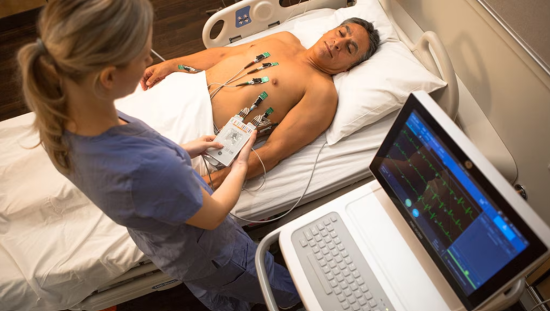
The absence of ST-segment abnormalities alone does not a "good" ECG stress test make.
By contrast, exercise testing is known to generate numerous variables, values, and metrics—well beyond ECG waveforms alone, and well after the exercise itself ends. This, you might say, amounts to a deluge of data.
Sorting through that deluge is necessary to paint a comprehensive picture of cardiovascular health during a heart stress test. With fuller insights, clinicians can better stratify a patient's risk and assess their prognosis and care.
But, how can physicians keep up with so many values? It starts by knowing what they are—and by expanding the scope of stress test evaluation to account for more than just exercise performance. Factors relative to recovery and risk are critical in determining overall cardiac fitness.
Here are values to consider before assuming a good ECG stress test is, in fact, good.
Heart Rate Recovery
Heart rate recovery is the measure of how quickly the heart rate goes back down to baseline levels after exercise is over. Typically, clinicians can measure this by subtracting the beats per minute exactly one minute after exercise termination from the heart rate beats per minute at peak exercise.
When the heart rate is slow to recover—with a number below 13, for example—it can be an indication of problems associated with the parasympathetic nervous system's reactivation process.1 This can be seen with heart failure patients.
Moreover, a low measurement can be a predictor of mortality. For example, a seminal study from 1999 found that an abnormally low heart rate doubled the risk of death over six years.1 Others have found similar results, indicating the prognostic value of this potentially underused metric.
Ventricular Ectopy
The presence of so-called "high-grade" premature ventricular ectopy can inform cardiovascular mortality in a way similar to heart rate recovery—meaning that this evaluation offers the most insight during recovery but not the actual exercise.
One paper in the Journal of the American College of Cardiology found that such ectopic events produced a hazard ratio of 1.82 for cardiovascular mortality. "High-grade," they explain, occurs when ectopic episodes are frequent (more than 10 per minute), R-on-T, multifocal, or recurring at a rate of two or more subsequently.2
Importantly, this value was prognostically beneficial only during the recovery period: High-grade events that happened during the physical activity did not achieve that same increased risk. This, again, emphasizes that exercise testing requires a fuller scope than the test itself. Measures of recovery are critical and can go under the radar when only surface-level metrics are analyzed.
Metabolic Equivalents (METS)
The METS value helps to quantify exercise capacity by way of oxygen uptake, with one advantage being its ability to produce a common metric (the equivalent) no matter the test or protocol.
It's known to be useful for predicting cardiac events after myocardial infarction, including on the cycle and treadmill. Generally, a calculation of less than 5 has been associated with poor prognosis, whereas achieving > 8 METs is associated with lower relative risk of death independent of other risk factors such as hypertension, diabetes, smoking, elevated body mass index (>30), elevated cholesterol (>220mg/dl), or comorbid chronic obstructive airways disease.3
Rate Pressure Product (RPP)
Also known as the Double Product, the RPP multiples the systolic blood pressure with the heart rate so that clinicians can better understand myocardial oxygen demand. Its prognostic value can be gleaned by calculating the maximum RPP at designated intervals throughout the exercise test. An RPP above 20,000 mmHg per minute is considered healthy, whereas anything below 16,000 mmHg is deemed insufficient.4
Poor RPP has been found to be a helpful predictor for cardiac events, and potentially better than both heart rate reserve (HRR) and age-predicted maximum heart rate.5
To learn more about the power of the ECG in today's clinical landscape, browse our Diagnostic ECG Clinical Insights Center.
Chronotropic Response
The chronotropic response determines the heart rate reserve used and can be highly valuable for helping to predict concerns of coronary artery occlusions. One study identified a relationship between the two at a rate of more than 50%. Additionally, low values of this assessment have been linked to carotid atherosclerosis, acute MI, and mortality.
Generally, values below 80% indicate chronotropic incompetence. This threshold is modified to 62% when a patient is taking beta blockers. As with other measurements, it can involve some complicated arithmetic: 100 x (HRpeak-HRrest)/(200-age-HRrest)35.4
T-Wave Alternans
T-Wave alternans (TWA), which is observed as an alternating pattern in the morphology of cardiac repolarization, assesses how multiple factors such as timing and amplitude change between consecutive beats on the ECG reading. It examines not just the T-wave but also the ST-segment. Because it can have a wide range of amplitudes, advanced systems that study the so-called "microvolts" of TWA have become helpful.6 Calculating the TWA requires a complex algorithm that can be automated with modern systems.4
This measurement can help identify patients at risk for ventricular arrhythmias, which commonly have alternans of the ST-segment and T-wave. TWA may also factor into the risk assessment for sudden cardiac death.6 The impacts of this measure during exercise testing have been studied both generally and with a lead-specific purview, of which lead five was shown to correlate most with mortality risk.7 TWA has also been found to have clinical utility in prognosticating death (cardiovascular and all-cause) when combined with heart rate recovery.8
Duke Treadmill Score (DTS)
For those using the treadmill during exercise testing, this ECG stress testing value can help to determine mortality across a low-, moderate-, and high-risk stratification among patients with known or suspected coronary artery disease. But because of its math-intensive process, it can be prone to errors if not cautiously calculated. Thankfully, many systems offer this as an automated measurement to support the user.
To determine DTS, multiply the maximum ST deviation (in millimeters) by five and the angina index by four. Then, subtract those numbers from the minutes of exercise duration. When the value is lower than -10, patients are at high risk.4 This highest tier of risk yields a prognostic five-year survival of just 65%.9
Understanding the Nuances of ECG Stress Tests
The stress testing data points that feed into cardiovascular health and function are nuanced and, at times, highly complex. They do, however, yield valuable prognostic information, which is invaluable in determining treatment and management of patients. In order to develop care plans more confidently, clinicians must account for them all. This takes time, effort, and resources—and likely isn't something practitioners can learn quickly.
That said, by familiarizing themselves with these prognostic variables, cardiologists can get a good start toward understanding the grey areas of everyday exercise testing. When complexities such as DTS, METS, and other metrics are considered, you might find far fewer "good" stress tests and many more cases that deserve a closer look.
Resources:
1. Cole C, Blackstone E, Pashkow F, et al. Heart-rate recovery immediately after exercise as a predictor of mortality. New England Journal of Medicine. 1999; 341:1351-7. https://www.nejm.org/doi/full/10.1056/NEJM199910283411804
2. Refaat MM, Gharios C, Moorthy MV, et al. Exercise-induced ventricular ectopy and cardiovascular mortality. Journal of the American College of Cardiology. https://www.acc.org/latest-in-cardiology/journal-scans/2021/11/30/19/50/exercise-induced-ventricular-ectopy. Accessed December 1, 2022.
3. Gibbons RJ, Balady GJ, Bricker JT, et al. American College of Cardiology/American Heart Association Task Force on Practice Guidelines. Committee to Update the 1997 Exercise Testing Guidelines. ACC/AHA 2002 guideline update for exercise testing: summary article. A report of the American College of Cardiology/American Heart Association Task Force on Practice Guidelines (Committee to Update the 1997 Exercise Testing Guidelines). Journal of the American College of Cardiology. 2002;40(8):1531-40. https://doi.org/10.1016/S0735-1097(02)02164-2.
4. Physician's Guide to GE Stress Systems. GE Healthcare. 2051167-002 ENG.
5. Whitman M, Jenkins C. Rate pressure product, age predicted maximum heart rate or heart rate reserve. Which one better predicts cardiovascular events following exercise or echocardiography? American Journal of Cardiovascular Disease. 2021;11(4):450-457. https://www.ncbi.nlm.nih.gov/pmc/articles/PMC8449196/
6. Narayan S. T wave (repolarization) alternans: overview of technical aspects and clinical applications, UpToDate.com. https://www.uptodate.com/contents/t-wave-repolarization-alternans-overview-of-technical-aspects-and-clinical-applications. Accessed December 2, 2022.
7. Leino J, Verrier RL, Minkkinen M, et al. Importance of regional specificity of T-wave alternans in assessing risk for cardiovascular mortality and sudden cardiac death during routine exercise testing. Heart Rhythm. 2011;8(3):385-390. https://doi.org/10.1016/j.hrthm.2010.11.004
8. Leino J, Minkkinen M, Nieminen T, et al. Combined assessment of heart rate recovery and T-wave alternans during routine exercise testing improves prediction of total and cardiovascular mortality: the Finnish Cardiovascular Study. Heart Rhythm. 2009;6(12):1765-1771. https://doi.org/10.1016/j.hrthm.2009.08.015
9. Duke treadmill score. Healio.com. https://www.healio.com/cardiology/learn-the-heart/cardiology-review/topic-reviews/duke-treadmill-score. Accessed December 1, 2022.


