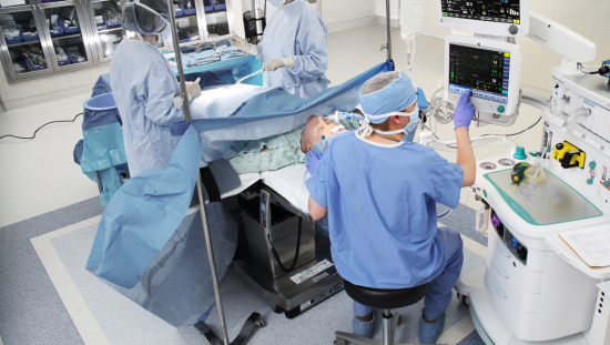
Many barriers exist to adopting LPV protocols, and these include a need to educate anesthesiologists on the benefits of adhering to LPV practices. This has prompted the development of automated anesthesia management using software algorithms to better anticipate patient ventilator needs during surgery.
Barriers to Implementing lung protective ventilation (LPV)
LPV has been proven to help reduce PPCs in both patients at high risk, as is the case in patients with acute respiratory distress syndrome (ARDS) and patients with perceived normal baseline lung function.1 Over the years, anesthesiologists have implemented LPV by adjusting tidal volume and positive end-expiratory pressure (PEEP) based on a handful of clinical parameters — weight, gender, surgical time, and preexisting conditions to name a few.
Although these parameters are helpful in determining LPV for surgical patients, they are not without their own limitations, ranging from guessing ideal body weight at the bedside to a lack of an institutional LPV protocol.
Clinical guesswork
LPV consists of using lower tidal volumes and proper use of PEEP, but even with adherence to guidelines, there remains room for error. An example of this is calculating tidal volume based on predicted body weight. Guidelines state to use predicted body weight (PBW), which is calculated by using height and gender to determine tidal volume, even though studies show this objective measurement isn’t always used.
A recent retrospective review, evaluating risk factors for excessive tidal volumes, explored reasons why this may be the case. Researchers found patients who were obese were at higher risk of PPCs due to a high tidal volume.2 The researchers hypothesized that this was caused by clinicians using actual body weight instead of predicted body weight (PBW), which is better correlated to lung size, when calculating the tidal volume setting for the ventilator.
By relying on clinicians to make an educated guess on someone’s PBW rather than calculating PBW using height and gender, this educated guess can lead to bias and error in tidal volume calculations that may lead to volutrauma.
Another reason a higher tidal volume may be used with an obese patient is that the patient’s actual weight is used to calculate LPV settings rather than calculating the PBW. This may be done in error or due to clinician routine practices.
Researchers also note that female patients were more likely to not receive proper LPV and were at higher risk of PPCs, citing a female PBW to be overestimated by the clinician more often than male patients.2
Clinician knowledge
Another barrier to implementing proper LPV is clinician knowledge. Clinicians tend to have a handful of perceptions when lowering tidal volume in order to achieve LPV and reduce PPCs. Nearly half (40%) are concerned about atelectasis caused by too low a tidal volume, while 36% are concerned about hypoxia and acidosis.3
Another barrier cited by 24% of anesthesiologists surveyed is the lack of an LPV protocol provided or implemented by the surgical institution to combat personal bias.3
When anesthesiologists have been trained in LPV, they tend to apply lower tidal volumes and use PEEP more frequently. As with all clinical advances and discoveries, there is an education period required before it is widely adopted as a clinical best practice.
The adoption of LPV strategies is no different, and this lack of education was illustrated in a 2018 questionnaire of anesthesiologists.4 Those who were formally trained and understood the importance of LPV were more likely to use it than their colleagues who had less knowledge, highlighting the need for proper and continuing education when it comes to preventing PPCs
Improving LPV efforts with automated anesthesia management
While it’s clear the current methods for creating LPV protocols for patients are helpful in reducing PPCs, there is much room for improvement — namely in reducing errors dependent on subjective criteria from the clinician. This has led to the development of many digital models, which use more detailed and accurate physiology-based parameters to determine ventilator settings.
These complex algorithms take into account very specific parameters for each patient. The focus of these algorithms has been centered around respiratory and ventilator mechanics as well as acid-base balance of the patient.5 Rather than focusing on statistical parameters like gender, weight, and surgical time, certain software models factored in the following variables:
- Respiratory drive
- Pharmacokinetics of propofol
- Acid-base homeostasis
- Ventilator mechanics
- Lung and respiratory muscle mechanics
When this model was applied in several simulations, researchers concluded the automated model focused on lung and diaphragm protection during ventilation. Based on these simulations, this automated anesthesia model was robust. Even though more research and clinical application was needed, it showed promise in reducing PPCs in surgical patients.
Perioperative clinical information systems: Bringing LPV digital solutions together
If the future of LPV to reduce PPCs is dependent on using automated tools, then creating platforms to combine the expertise of the clinical staff with advanced algorithms for automated LPV is vital.
Using comprehensive and integrated information systems and platforms allows clinicians to review lab and radiology data along with ventilator and infusion settings in real time. These systems can improve communication outside of the OR by integrating with the host electronic medical record system — meaning the patient’s records are up-to-date before they enter the PACU. With comprehensive information tools, multisystem algorithms can work in tandem with clinicians to reach the highest standards for lung protective ventilation.
References
- LAS VEGAS investigators. 2017. Epidemiology, practice of ventilation and outcome for patients at increased risk of postoperative pulmonary complications. Eur K Aneasthesiol. 34:492-507.
- Koaw, CY et al. 2020. Risk factors for excessive tidal volumes delivered during intraoperative mechanical ventilation, a retrospective study. Int J Physiol Pathophysiol Pharmacol. 12(2). 51-57.
- Kidane B, Choi S, Fortin D, O’Hare T, Nicolaou G, Badner NH, Inculet RI, Slinger P, Malthaner RA. Use of lung-protective strategies during one-lung ventilation surgery: a multi-institutional survey. Ann Transl Med 2018;6(13):269. doi: 10.21037/atm.2018.06.02.
- Kim, SH et al. 2018. Application of intraoperative lung protective ventilation varies in accordance with the knowledge of anaesthesiologists: a single-centre questionnaire study and a retrospective observational study. BMC Anesthesiology. 18(33).
- Zhang, B. et al. 2020. A physiology-based mathematical model for the selection of appropriate ventilator controls for lung and diaphragm protection. Journal of Clinical Monitoring and Computing. https://doi.org/10.1007/s10877-020-00479-x.
About GE HealthCare Technologies, Inc.
GE HealthCare is a leading global medical technology, pharmaceutical diagnostics, and digital solutions innovator, dedicated to providing integrated solutions, services, and data analytics to make hospitals more efficient, clinicians more effective, therapies more precise, and patients healthier and happier. Serving patients and providers for more than 100 years, GE HealthCare is advancing personalized, connected, and compassionate care, while simplifying the patient’s journey across the care pathway. Together our Imaging, Ultrasound, Patient Care Solutions, and Pharmaceutical Diagnostics businesses help improve patient care from diagnosis, to therapy, to monitoring. We are a $19.6 billion business with 51,000 colleagues working to create a world where healthcare has no limits. Follow us on LinkedIn, X (formerly Twitter), and Insights for the latest news, or visit our website https://www.gehealthcare.com/ for more information.
Products mentioned in the material may be subject to government regulations and may not be available in all countries. Shipment and effective sale can only occur after approval from the regulator. Please check with your local GE HealthCare representative for details. © 2025 GE HealthCare. GE is a trademark of General Electric Company used under trademark license. July 2025 JB16471XX


