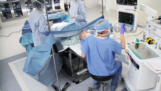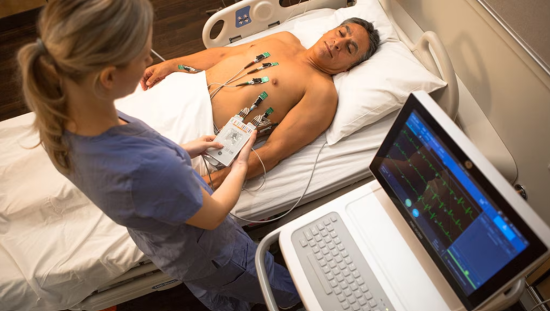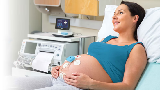
Every year, millions of people undergo surgical procedures. Patients are typically closely monitored during the perioperative period by the surgical team, but we know as patients enter the postoperative period – monitoring gradually and continuously declines.
While the expectation is for patients to improve and need less monitoring, the truth is postoperative complications and deaths occur at an alarming rate. According to an analysis published in The Lancet, 4.2 million people die worldwide within the 30-day window after surgery. This makes postoperative death a major cause of death and an issue worth improving upon.[1]
One way to attempt to improve postoperative outcomes is by improving postoperative vital sign monitoring. Patients on the general ward tend to slowly deteriorate with vital signs trending up or down into abnormal ranges over time rather than suddenly changing.[2] Additionally, interventions are time sensitive, and the sooner signs of deterioration are spotted, the better the outcomes.[3]
Rather than trying to monitor every vital sign continuously which would be extremely challenging, targeting the most predictive vital sign is a more achievable goal. Respiratory rate is the highest ranked vital sign in predicting clinical deterioration.[4],[5]
If a patient has respiratory compromise, they are 29 times more likely to die making respiratory status a critical component to desired outcomes in the postoperative period.[6]
Current systems used to monitor respiratory rate on a continuous basis are useful but are not without difficulties – especially when patients become ambulatory. Today we’re going to cover limitations related to both capnography and traditional impedance respiratory rate monitoring and discuss new technology that addresses many of these limitations.
Capnography – An Unwanted Tether
Capnography allows clinicians to assess ventilation status by monitoring changes to end-tidal CO2. Using capnography has been proven to improve patient safety and there is mounting evidence of the benefits of using continuous capnography monitoring.[7]
Identifying respiratory depression before it is detected on clinical exam and even before oxygen desaturation are two benefits of continuous capnography monitoring.7
Continuous capnography monitoring is a great option for patients in the ICU who are otherwise bedbound with limited ambulation. However, when patients enter the general ward and are encouraged to get up and move around postoperatively - capnography monitoring can be cumbersome.7
Capnography requires the use of a nasal cannula which can be uncomfortable to patients resulting in poor patient compliance. Also, because the patient is tethered to the bed with tubing, postoperative ambulation may be delayed.7
Additionally, capnography can require frequent recalibration which requires time from the clinician that could be spent elsewhere.
Traditional Impedance Monitoring – Too Much Noise
Impedance pneumography or simply impedance monitoring is another way to monitor respiratory rate that does not involve being tethered to a nasal cannula. Impedance monitoring uses thoracic electrical signals, traditionally with only one vector to monitor respiration rate.[8]
Even though it’s less cumbersome compared to continuous capnography monitoring, traditional impedance monitoring has been shown to be less than perfect when it comes accuracy.8
Artifact, or ‘noise’ from the impedance signal can contribute to inaccuracies in monitoring. Patient movement, lead placement (which often uses ECG electrode placements), and electrical signal noise generated by the patient’s cardiac rhythm can all contribute to artifact.
Proper lead placement is critical to accurate impedance monitoring and some studies show ideal lead placement locations can change based on patient position and how the patient breathes. An example being if they use their abdomen or their chest primarily with respiration.[9]
Impedance monitoring also relies on algorithms to evaluate respiratory rate and studies have found many algorithms currently being used have large margins of error.8
The inaccuracies of impedance monitoring contribute to false alarms or respiratory decline being missed by clinicians.8
Improving Continuous Respiratory Rate Monitoring
If accuracy of impedance monitoring could be increased, this would be a great way to improve continuous monitoring of respirations while still providing ample movement and freedom for patients.
One way this can be done is by monitoring two channels, or vectors, instead of just one. By adding an electrode and using a dual vector approach, accuracy can be improved with little to no added effort by nurses or patients.
The dual vector approach is comprised of three electrode applications versus two where placement has been optimized for respiration rate vs. relying on cardiac lead placements. A recent study has shown by adding an electrode and monitoring two impedance signals at the same time, this new technology has a greater than 99% measurement accuracy when compared to capnography.2
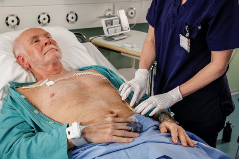
The accuracy of impedance monitoring not only depends on lead placement and signal quality – it greatly depends on the accuracy of the algorithm being used. Using highly effective algorithms to reduce false alarms and clean up any artifact from the sensors can help improve accuracy, providing clinicians with confidence in the technology and helping to speed adoption.
Algorithms must also be able to adjust for patient conditions and utilize intelligence to account for patient movement, shivering, or other external factors.
The TruSignal RRdv™ algorithm used by GE HealthCare’s Portrait Mobile device monitors two respiration vectors simultaneously, enabling the system to accurately measure respiration rate even if one vector has signal noise or low amplitude. By combining this with frequency analysis and dynamic delays the TruSignal RRdv technology can enable clinicians to continuously monitor their patients, even during ambulation, without the fear of excessive alarms.
As more research shows the importance of continuous respiratory monitoring outside of the surgical and ICU settings to help improve patient outcomes, technology must adapt to support ambulatory patients. This means improving accuracy of respiratory monitoring without compromising movement or safety and introducing dual-vector impedance monitoring is a great way to achieve this.
Summary
- Continuous vital sign monitoring can help identify patient deterioration postoperatively2
- Monitoring postoperative patients is challenging once patients start ambulating2
- Capnography is accurate but uncomfortable for patients and hinders ambulation due to nasal canula
- Traditional impedance respiratory monitoring allows for more mobility, but struggles with respiratory monitoring accuracy
- Dual-vector impedance monitoring increases accuracy by optimizing respiration rate signal and helping to minimize false alarms
- Combined with intelligent algorithms, dual-vector impedance monitors like GE HealthCare’s Portrait Mobile monitoring solution has the potential to improve accuracy while encouraging mobility with a sleek wearable, wireless device.
References
[1] Nepogodiev, D, Martin, J, Biccard, B, Makupe, A, & Bhangu, A. (2019). Global burden of postoperative death. The Lancet, 393 (10170). P401.
[2] Jarvela, K, Takala, P, Michard, F, & Vikatmaa, L. (2021). Clinical evaluation of a wearable sensor for mobile monitoring of respiratory rate on hospital wards. Journal of Clinical Monitoring and Computing. 36. 81-86.
[3] Mebazaa, A., et al. (2016). Designing phase 3 sepsis trials: application of learned experiences from critical care trials in acute heart failure. J Intensive Care. 4, 24.
[4] Michard, F., Sessler, D. (2018). Ward monitoring 3.0. Br J Anaesth, 121(5):999-1001.
[5] Michard, F, et al. (2022). Association Between Mean Arterial Pressure and Acute Kidney Injury and a Composite of
Myocardial Injury and Mortality in Postoperative Critically Ill Patients: A Retrospective Cohort Analysis. BJA Open. 1(c): 1000002.
[6] Kelley SD, SA, Agarwal S, Parikh N, Erslon M, Morris P. (2012). Respiratory insufficiency, arrest and failure among medical patients on the general care floor. Crit Care Med. 40(12):764.
[7] Thach, L et al. (2017). Continuous pulse oximetry and capnography monitoring for postoperative respiratory depression and adverse events: a systemic review and meta-analysis. Anesthesia and Analgesia. 125 (6). 2019-2029.
[8] Charlton, P et al. (2021). An impedance pneumography signal quality index: design, assessment and application to respiratory rate monitoring. Biomed Signal Process Control. 65. 102339.
[9] Bawua, L, Miaskowski, C, Hu, X, Rodway, G. & Pelter, M. (2021). A review of the literature on the accuracy, strengths, and limitations of visual, thoracic impedance, and electrocardiographic methods used to measure respiratory rate in hospitalized patients. Ann Noninvasive Electrocardiology. 26(5). E12885.
Not all products or features are available in all markets.

