Too many patients continue presenting to the post anesthesia care unit (PACU) with residual paralysis, despite the increasing availability and accuracy of anesthesia monitoring technology.1
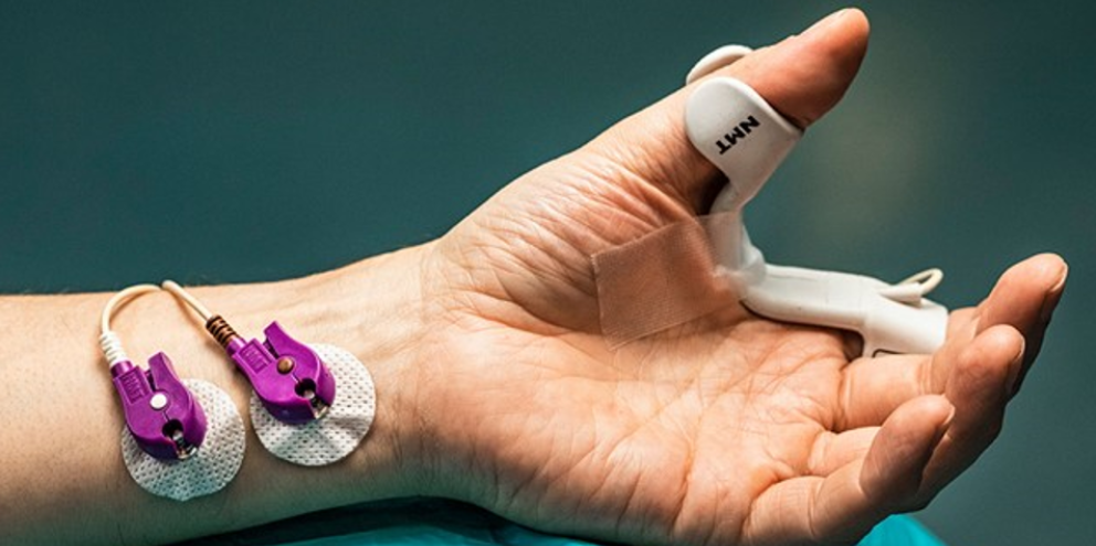
Acceleromyography (AMG) and electromyography (EMG) are the two forms of quantitative neuromuscular transmission monitoring. EMG monitoring has come a long way and is the newest technology for providing accurate, reliable train-of-four (TOF) measures.
Neuromuscular blocking agents (NMBAs) have variable durations of action, so there's no set range of time in which clinicians can guarantee a patient has achieved that range of recovery. In addition, having an accurate TOF reading from a quantitative source rather than qualitative assessment helps avoid residual paralysis to improve patient outcomes.1
"Unless you monitor, you don't know," said Stuart Grant, MD, chief of regional anesthesia at the University of North Carolina Chapel Hill and a co-author of the ASA guidelines. "If you want to be the best provider, you have to monitor."
Quantitative monitoring solutions to improve patient safety
AMG monitoring has been the go-to choice for the past few decades, but easy-to-use EMG machines have become available over the past few years. EMG provides advantages compared with AMG, especially for robotic and laparoscopic surgery. AMG characterizes movement by measuring acceleration of the limb (indirect measurement), whereas EMG measures the electrical activity of the muscle (direct measurement).
AMG monitoring
AMG measures a muscle contraction in the adductor pollicis in response to stimuli at the ulnar nerve to determine the TOF ratio. AMG has plenty of benefits over not monitoring at all, though it also has its downfalls; it is less precise when measuring the train-of-four (TOF) ratio, which is the key indicator used for timing extubation.
These machines can overestimate the TOF ratio by 0.1. Studies have shown that AMG monitoring can produce TOF ratios more than 1.0, and can overestimate a TOF ratio by at least 0.15.2 That makes it difficult to know for sure if a patient measuring a TOF ratio of 0.9 (the ratio at which patients are considered to have no residual paralysis) with AMG has fully recovered.3 Clinicians need to normalize the TOF ratio before a procedure to ensure accuracy when using an AMG monitor.
"If you do not standardize AMG beforehand, you really want a ratio of 1.0 or above to guarantee recovery," Dr. Grant said.
One limitation of AMG for certain surgeries is that it requires free movement of the thumb to get an accurate reading. That presents a challenge to continuously monitoring a patient during a surgery where the patient's hands are tucked, such as robotic surgery.3 To accurately get a reading, the clinician can measure the TOF ratio at the end of surgery when the patient's hand can be freed.
EMG monitoring
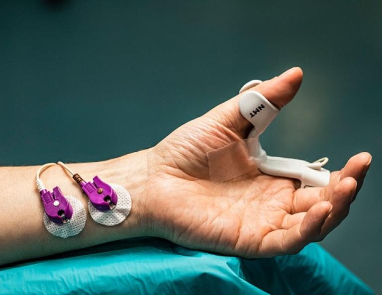
By contrast, EMG measures the electrical signal in the muscle, providing a reading at the source of action for the NMBAs. There's less potential for noise in the signal, and there are more precise measurements when compared to measuring the muscle twitch with AMG.3
One of the major advantages of EMG is that it doesn't require free thumb movement. Increasingly, surgeries are performed robotically or laparoscopically, which requires a patient's hands to be tucked closely to the body. EMG can be used even when the thumbs are tucked.
"It doesn't matter if your thumb is open or your hands are free, EMG is going to give an accurate measurement," Dr. Grant said. "The EMG gives freedom in patient positioning. In our practice where robotic surgeries are increasing, EMG has made a big difference."
Applying EMG monitoring in practice
Monitoring patients throughout surgery can improve patient safety, provide valuable information for reversal, and improve efficiency in the operating room. The ultimate goal of anesthesia monitoring is to avoid residual paralysis in the PACU.
Residual paralysis can lead to complications such as upper airway obstruction, pneumonia, and a prolonged stay in the PACU.1 Dr. Grant pointed out that aspiration and pneumonia are two of the more common complications of inadequate recovery. However, pneumonia often develops days later, making it harder to draw a connection to the operating room. In rare cases, patients who don't adequately recover need reintubation, which leads to an ICU stay and more risks and costs for the patient.
Quantitative monitoring can affect multiple decision points throughout surgery to help ensure a smoother experience for the clinician and patient.
- Monitor depth of neuromuscular block throughout surgery. Continuous monitoring is part of the GE HealthCare Adequacy of Anesthesia approach to ensure appropriate dosing and safety; it measures the patient's depth of block during surgery. Real-time monitoring during surgery allows for redosing as needed if the patient starts to become more conscious or responsive. According to Dr. Grant's observation, quantitative monitoring throughout a procedure helps predict how quickly a patient will become fully conscious and recover from the anesthetic agent. Access to quantitative monitoring allows the proactive dosing of reversal agents to help patients safely emerge from the effects of NMBAs timely.
- Accurately dose reversal agents. Patients metabolize reversal agents differently based on age, weight, gender, and other factors. Appropriate dosing depends on accurate assessment of the depth of neuromuscular block, which can be achieved with EMG monitoring.1 Accurate dosing may reduce the liberal use of reversal agents and can improve efficiency to time of recovery, enabling patients to be moved to the PACU without the risk of unknown residual paralysis.
- Appropriately time extubation. The ASA guidelines recommend confirming a TOF ratio of >0.9 with quantitative monitoring before extubation. EMG monitoring provides the most accurate readings without having to determine a baseline ratio before surgery.3
Dr. Grant has found that quantitatively monitoring patients throughout surgery allows him to predict how patients will respond to reversal agents and how quickly. "When a patient's fully reversed, there's no such thing as a slow recovery," he said. "That means there's less time in the OR, patients recover promptly and with fewer issues, and you get a better flow to your practice."
EMG monitors continue to set the standard for quantitative neuromuscular monitoring, with application in a variety of clinical settings. Newer machines have easy-to-use interfaces, disposable sensors with easy placement instructions, and clinical decision-making support features built in. Quantitative monitoring provides clinicians with confidence in their readings, offering a precise and reliable method to monitor patients. This contributes to improving patient safety and clinical efficiency by helping patients fully recover and smoothly emerge from anesthesia in a timely manner.
Resources:
1. Thilen SR, Weigel WA, Todd MM, et al. 2023 American Society of Anesthesiologists practice guidelines for monitoring and antagonism of neuromuscular blockade: A report by the American Society of Anesthesiologists task force on neuromuscular blockade. Anesthesiology. 2023;138(1):13-41. doi:10.1097/ALN.0000000000004379.
2. Liang S, Phillips S, Stewart PA. An ipsilateral comparison of acceleromyography and electromyography during recovery from nondepolarizing neuromuscular block under general anesthesia in humans. Anesthesia & Analgesia. August 2013; 117(2):373-379. https://journals.lww.com/anesthesia-analgesia/fulltext/2013/08000/an_ipsilateral_comparison_of_acceleromyography_and.14.aspx. Accessed October 9, 2023.
3. Bowdle A, Bussey L, Michaelsen K, et al. A comparison of a prototype electromyograph vs. a mechanomyograph and an acceleromyograph for assessment of neuromuscular blockade. Anaesthesia. 2020;75(2):187-195. doi:10.1111/anae.14872

