These deaths can be due to many elements and perioperative strokes are one of them. Up to 6% of cardiac surgical patients experience a stroke during surgery, and this is associated with an increased length of stay in the hospital and increased mortality.[1]
A compounding issue is these strokes are not always detected and can be covert with minimal or no symptoms present. This is problematic because covert strokes increase the risk of an acute ischemic stroke in the future.[2]
Low cerebral oxygenation can precipitate a stroke, and unfortunately, happens frequently in certain types of surgery. Over 30% of high-risk cardiac surgery patients in the intensive care unit (ICU) experience cerebral desaturation which is defined as a greater than 20% drop from baseline.[3]
Of those patients, they aren’t just experiencing one event either – most have an average of five desaturation events postoperatively.4
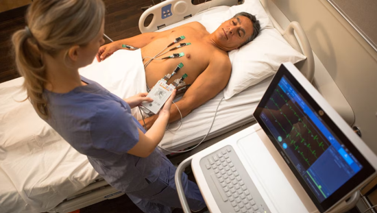
Being able to monitor cerebral saturation as well as overall hemodynamic stability is crucial in the perioperative period because of these risks. With close monitoring, clinicians can hopefully detect changes and potential desaturation and intervene in a timely manner.
ECMO Complicates Monitoring
Pulse oximetry is commonly used in the operating room (OR) and ICU to monitor oxygenation, but cannot monitor specific organs or tissue. This becomes an issue when certain procedures are done in the OR like extracorporeal membrane oxygenation (ECMO), which is commonly used in cardiac surgery.
ECMO is a procedure that removes carbon dioxide from the blood and then introduces oxygenated blood back into the body. This is done in a machine outside of the body with the goal being to allow the heart and lungs to rest during a time of illness or recovery.[4]
Risks related to ECMO include infection, vascular complications, and limb ischemia.[5] Another complication related to ECMO is the risk of dual circulation or “North South Syndrome”.6
This occurs when there is an imbalance of oxygenated blood where one area of the body is adequately oxygenated, and another area is hypoxic. North South Syndrome can cause low cerebral partial pressures of oxygen.6
This means a pulse oximeter may report a normal oxygenation reading but in reality, one area is really struggling to maintain proper oxygen levels.
Cerebral desaturation from North South Syndrome can be fatal and highlights the importance of being able to monitor oxygenation of various organ systems in addition to overall oxygenation.6
What is Near Infrared Spectroscopy and How Does it Work?
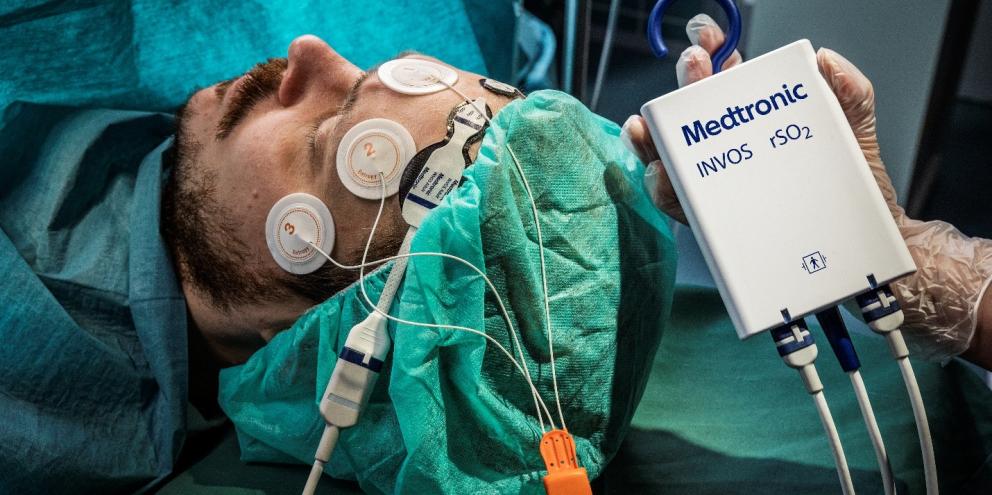
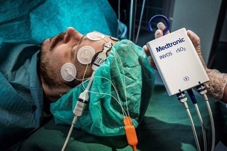
Near infrared spectroscopy (NIRS) is a monitoring device that provides accurate oxygenation information. NIRS evaluates the balance of oxygen supply versus consumption in the brain and other organ systems.[6]
As the name implies, near-infrared spectroscopy uses near-infrared light to determine oxygenation levels in brain tissue. This is done by using a light source fixed to the head that transmits light through the skin, skull, connective tissues, and brain.
A detector then quantifies the light to measure cerebral oxygen saturation in real time.[7]
Benefits of Near Infrared Spectroscopy
With NIRS, clinicians can access cerebral/somatic monitoring in real time to visualize regional tissue oxygen extraction and utilization. NIRS provides oxygen saturation from vascular beds to both individually and collectively assess organs.[8]
Earlier intervention may be possible with NIRS due to clinicians being alerted to ischemic threats in the cerebral and peripheral circulatory systems before they become problematic.9
NIRS has been proven to be an accurate and effective measure of cerebral oxygenation. Studies have found that when NIRS is utilized, there is a drastic reduction in major organ morbidity or mortality in patients undergoing coronary artery bypass surgery. This is due to NIRS helping patients avoid major cerebral desaturation.[9]
Other studies have found patients require fewer intraoperative red blood cell transfusions and have shorter durations of ventilation when NIRS is used.[10],[11]
When patients go to the ICU postoperatively, NIRS monitoring can help reduce the amount of time patients are experiencing cerebral desaturation events by 48% compared to those without NIRS monitoring.[12]
NIRS Use in Nonsurgical Settings
While NIRS has been proven helpful in the intraoperative and immediate postoperative setting, there may be an additional benefit for NIRS monitoring in the ICU setting. Over half (60%) of cardiac surgery patients experience a desaturation event in the four hours post-surgery.[13]
When surveyed, cardiovascular ICU nurses taking care of these patients reported NIRS devices to be easy to use and helpful to monitor hemodynamics. Half of nurses surveyed felt NIRS monitoring positively impacted patient care.14
Nevertheless, more research is needed to further determine the role NIRS monitoring may play in the ICU. Due to its noninvasive method, it could be a useful, low-risk tool to include in ICU care.
Summary
- Cerebral desaturation in the OR and postoperative period are a major concern, especially in high-risk cardiac surgeries and critically-ill patients
- Monitoring cerebral and other organ tissue oxygenation provides valuable data that help enable clinicians reduce the risk of desaturation periods
- NIRS provides a noninvasive method to monitor oxygenation and oxygen use in real time and reduce desaturation events
- While NIRS has been proven effective in the surgical and immediately postoperative setting, there may be benefits to using in the ICU, but more research is needed to fully understand benefits in this setting
References:
[1] Raffa GM, Agnello F, Occhipinti G, et al. (2019). Neurological complications after cardiac surgery: a retrospective case-control study of risk factors and outcome. J Cardiothorac Surg. 2019;14(1):23.
[2] Meinel, T et al. (2020). Covert brain infarction. AHA Journals: Stroke. 51(8):2597-2606.
[3] Deschamps A, et al. (2016). Cerebral Oximetry Monitoring to Maintain Normal Cerebral Oxygen Saturation during High -risk Cardiac Surgery: A Randomized Controlled Feasibility Trial. Anesthesiology, 124.
[4] Mayo Clinic. (2023). Extracorporeal membrane oxygenation. Patient Care & Health Information. https://www.mayoclinic.org/tests-procedures/ecmo/about/pac-20484615
[5] Pillai, A et al. (2018). Management of vascular complications of extra-corporeal membrane oxygenation. Cardiovascular Diagnostic Therapy, 8(3):372-377.
[6] Vretzakis, G. (2013). Monitoring of brain oxygen saturation (INVOS) in a protocol to direct blood transfusions during cardiac surgery: a prospective randomized clinical trial. J Cardiothorac Surg, 8:145.
[7] Takegawa, R et al. (2020). Near-Infrared Spectroscopy Assessments of Regional Cerebral Oxygen Saturation for the Prediction of Clinical Outcomes in Patients With Cardiac Arrest: A Review of Clinical Impact, Evolution, and Future Directions. Frontiers in Medicine, 7:587930.
[8] Marin T and Moore J. (2011). Understanding Near-Infrared Spectroscopy J. Adv Neonatal Care, 11(6):382-8
[9] Murkin, J et al. (2007). Monitoring brain oxygen saturation during coronary bypass surgery: a randomized, prospective study. Anesth Analg, 104(1):51-8.
[10] Lejus C, et al. (2015). A retrospective study about cerebral near-infrared spectroscopy monitoring during paediatric cardiac surgery and intra-operative patient blood management. Anaesth Crit Care Pain Med..22.
[11] Goldman SM, et al. (2004). Optimizing Intraoperative Cerebral Oxygen Delivery Using Noninvasive Cerebral Oximetry Decreases the Incidence of Stroke for Cardiac Surgical Patients. The Heart Surgery Forum, 5.
[12] Lei L, et al. (2017). Cerebral oximetry and postoperative delirium after cardiac surgery: a randomised, controlled trial. Anaesthesia. 72.
[13] Slater T, et al. (2017). Cerebral Perfusion Monitoring in Adult Patients Following Cardiac Surgery: An Observational Study. Contemporary Nurse.

