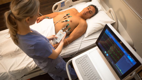
As experienced clinicians know, cardiac patients often present with wide QRS complexes, making it difficult to differentiate ventricular and supraventricular tachycardia. The case study below highlights the importance of monitoring more than one precordial ECG lead to detect and differentiate these cardiac arrhythmias.
Case study
In a 2010 white paper,1 Dr. Barbara Drew described a patient case that highlights the risks of monitoring using only one precordial lead.
The case study features a 62-year-old man who presented with a history of stable angina pectoris and recent cardiac catheterization showing stenotic lesions in the left anterior descending coronary artery was admitted to a telemetry unit with acute shortness of breath and near-syncope. Nursing responsibilities included monitoring the patient for arrhythmias and acute myocardial ischemic episodes in order to correlate electrocardiogram (ECG) changes with symptoms. However, this need to detect two different kinds of events posed a dilemma, as outlined henceforth.
It is understood that the best ECG lead for monitoring arrhythmias is V1.2,3 The patient’s symptoms were related to a wide QRS complex tachycardia, and V1 is capable of distinguishing ventricular tachycardia (VT) from supraventricular tachycardia (SVT) with aberrant conduction. However, the best ECG lead for monitoring acute ischemia related to the left anterior descending coronary artery is V3.4.5 Therefore, ideally, both V1 and V3 would be utilized in this patient. Unfortunately, as is typical, the available telemetry system allowed only for the monitoring of one precordial lead. Faced with two imperfect options, the nurse selected V1.
The patient went on to experience SVT with an aberrant ventricular conduction as noted by the QRS configuration in lead V1. The patient was unaware of any symptoms at that time, so the arrhythmia was not considered as the cause of his presenting symptoms. Approximately one hour later, the patient’s monitor alarm sounded and the rhythm strip showed a sustained wide QRS complex tachycardia at a rate of 188 beats per minute. The patient complained of shortness of breath and faintness. This time, it was clear that the arrhythmia was VT because of the QRS configuration in V1. A bolus of lidocaine was therefore administered, which terminated the tachycardia.
Implications for patient care
Choosing to monitor lead V1 here proved valuable for this patient because it provided ECG criteria that crucially distinguished between SVT and VT. Without these data, the nurse may have mistaken sustained tachycardia for SVT because the patient did not lose consciousness. Verapamil, a calcium channel blocker commonly used to resolve SVT, is not effective in VT. In fact, the drug is contraindicated in patients with VT because of negative inotropic effects and the potential to cause sudden hemodynamic deterioration and cardiac arrest.
Two standard 12-lead ECGs were recorded inadvertently (as part of technician training) just before the sustained VT. One showed an ischemic event with striking ST-segment elevation in leads V3 through V5. Of note, the ST segment in V1 did not change during this time, which explains why the ST monitor alarm was not triggered. This unexpected information indicated that the trigger for the patient’s malignant ventricular arrhythmia was acute ischemia. The patient was scheduled to undergo coronary bypass surgery the next day, and intravenous nitroglycerin was initiated with an order to titrate upward in the event of the recurrence of an ischemic episode.
The nurse in this case realized that this documentation of an ischemic episode was a fortunate accident—the ischemic episode was detected due to the two standard 12-lead ECGs, and would have been missed with routine cardiac monitoring. The nurse then faced the dilemma of how best to monitor the patient for both arrhythmias and ischemia during the 24 hours prior to surgery. Because the patient experienced silent ischemia, the nurse could not rely on symptoms of chest pain to detect acute ischemic episodes. Without such symptoms, the nurse also would not be prompted to record a “stat” 12-lead ECG in order to document further ischemic episodes.
In the present case, continued monitoring with V1 means that the titration of nitroglycerin must be based on guesswork. However, if the monitoring is changed to V3, any future episodes of wide QRS complex tachycardia would not contain the QRS criteria needed to distinguish between VT and SVT. In light of this conundrum, it is clear that what is needed is a telemetry system that enables the simultaneous monitoring of two precordial leads so that patients can be monitored for arrhythmias as well as ischemia.
- Drew BJ. Value of monitoring a second precordial lead for patients in a telemetry unit. GE HealthCare. 2010.
- Drew BJ. Bedside electrocardiographic monitoring: State of the art for the 1990s. Heart Lung. 1991;20(6):610–623. Accessed May 1, 2019.
- Wellens JJ, Bär FW, Lie KI. The value of the electrocardiogram in the differential diagnosis of a tachycardia from a widened QRS complex. Am J Med. 1978;64(1):27–33. Accessed May 1, 2019.
- Bush HS, Ferguson JJ 3rd, Angelini P, Willerson JT. Twelve-lead electrocardiographic evaluation of ischemia during percutaneous transluminal coronary angioplasty and its correlation with acute reocclusion. Am Heart J. 1991;121(6):1591–1599. Accessed May 1, 2019.
- Drew BJ, Tisdale LA. ST segment monitoring for coronary artery reocclusion following thrombolytic therapy and coronary angioplasty: identification of optimal bedside monitoring leads. Am J Crit Care. 1993;2(4):280–292. Accessed May 1, 2019.


